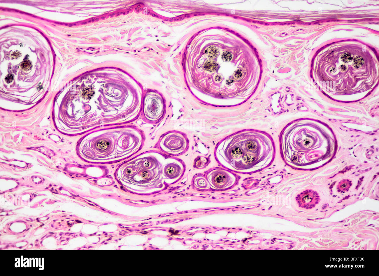
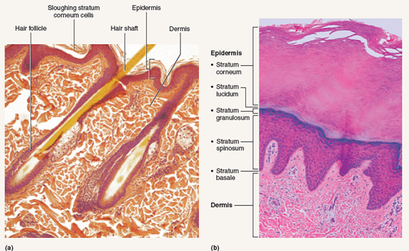
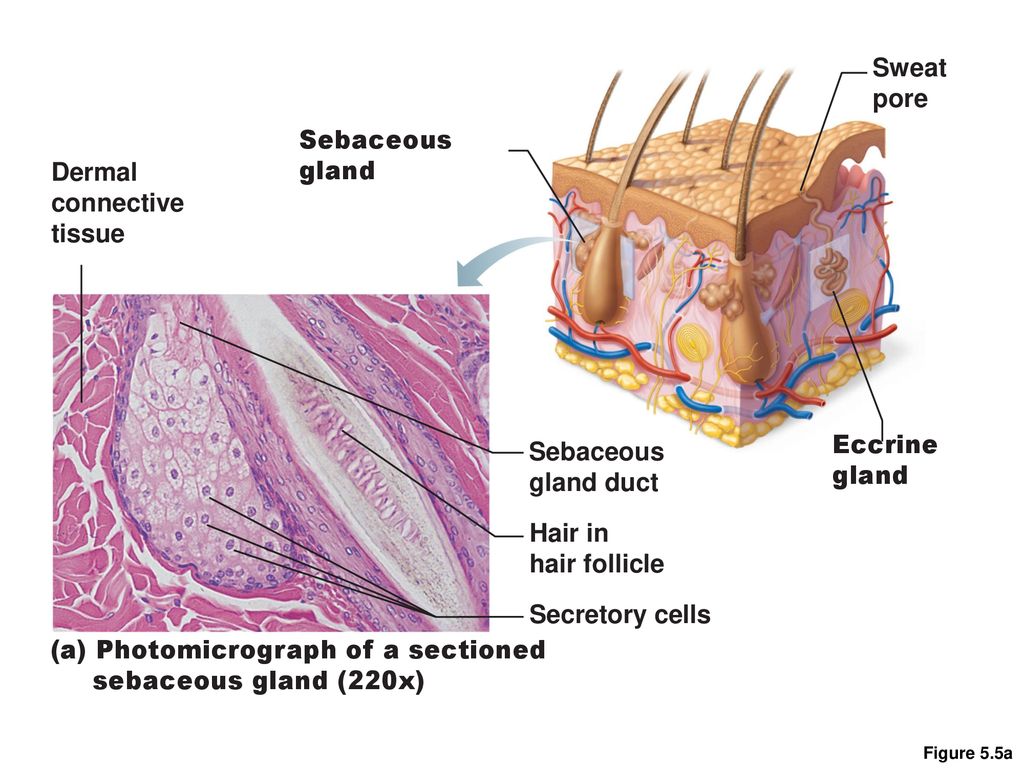
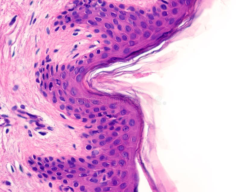
44 label the photomicrograph of thin skin.
Answered: Looking at the entire photomicrograph,… | bartleby Q: Label the internal structure to which the lines are pointing to A: -The human heart is the four-chambered muscular organ, formed and sized roughly like man's closed… Q: Skin - Model Drag the cursor over the labels to identify the parts Thick skin Thin skin 11 10 3 12… Label The Photomicrograph Of Thick Skin / Solved Label The ... The epidermis of thick skin has five layers: Thick skin · stratum basale (also known as s. Label the photomicrograph of thick skin. It has a fifth layer,. Start studying photomicrograph of the epidermal layer in thick skin. The outer layer of cells in this micrograph is the thinnest layer and. A few layers of cells that are .
Label The Photomicrograph Of Thick Skin - Faktor yang Label the photomicrograph of thick skin. 1 answer to label the photomicrograph of thin skin. The epidermis of thick skin has five layers: Hypodermis label the layers of the epidermis in thick skin in figure 7.2. A few layers of cells that are . Apocrine sweat gland label the photomicrograph in figure 7.4. Label the photomicrograph of thick skin.
Label the photomicrograph of thin skin.
Leaf Structure Under the Microscope Allow the nail polish about four hours to dry. Using a pair of tweezers, peel off a film (thin skin) from the surface of the leaf. Gently place the film onto a microscope slide and cover with a cover slip. Start with low power and increase to 100x (frequency of stoma can be counted at 100x) Record your observations. Anatomy, Skin (Integument), Epidermis - StatPearls - NCBI Bookshelf Skin is the largest organ in the body and covers the body's entire external surface. It is made up of three layers, the epidermis, dermis, and the hypodermis, all three of which vary significantly in their anatomy and function. The skin's structure is made up of an intricate network which serves as the body's initial barrier against pathogens, UV light, and chemicals, and mechanical injury ... photomicrograph of thick skin Diagram | Quizlet photomicrograph of thick skin Diagram | Quizlet photomicrograph of thick skin STUDY Learn Write Test PLAY Match Created by mckennawebber Terms in this set (7) epidermis (stratum corneum - stratum basale) ... stratum corneum ... stratum lucidum ... stratum granulosum ... stratum spinosum ... stratum basale ... dermis ... njordan6 mckennawebber
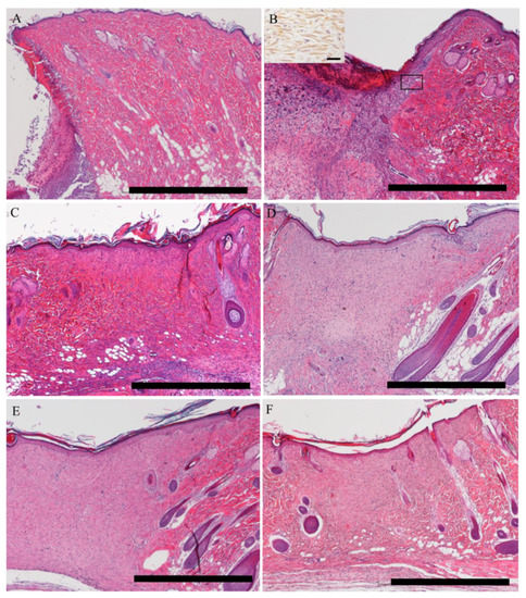
Label the photomicrograph of thin skin.. Photomicrograph of Thin Skin Quiz - PurposeGames.com This is an online quiz called Photomicrograph of Thin Skin There is a printable worksheet available for download here so you can take the quiz with pen and paper. Your Skills & Rank Total Points 0 Get started! Today's Rank -- 0 Today 's Points One of us! Game Points 5 You need to get 100% to score the 5 points available Actions Add to Playlist Label the photomicrograph in Figure 7.4. Examine a slide of hairy skin ... Label the layers of the skin on the diagram and the photograph. Be able to identify the layers on a microscope slide. Look at the skin slide under a microscope. a) Epidermis 1) Stratum corneum ii) Stratum lucidum 111) Stratum granulosum iv) Stratum... Posted one year ago Recent Questions in Basics of Statistics Q: Question : Question 31 points Label the photomicrograph of thin skin ... 31 points Label the photomicrograph of thin skin. Hair Follicle Hair Dermis Sebaceous gland Duct of sebaceous gland Reset zoom. Solution. 5 (1 Ratings ) Solved. Biology 2 Years Ago 77 Views. This Question has Been Answered! View Solution. Related Answers. unit 4 lab.docx - LAB Unit 4 EXERCISE 7: The Integumentary... FIGURE 7.2: Photomicrograph of the skin. epidermis (EPI-derm-is) • dermal papillae (puh-PILL-ee) • hypodermis (HY-poh- der-mis) • papillary (PAP-il-lary) layer of dermis • reticular layer of dermis 1. Dermal Papillae 2. Epidermis 3. Papillary layer of dermis 4. Reticular layer of dermis 5. Hypodermis
PDF Name the Condition Name the 4 layers of thin skin in both the cartoon and the photomicrograph. Name the 4 layers of thin skin in both the cartoon and the photomicrograph. •Name the Layers of skin and label the dermal papilla and dermis •Name the Layers of skin and label the dermal papilla Sebaceous Gland Label The Photomicrograph Of Thin Skin - Integumentary ... Label the photomicrograph of thin skin. Part a is a micrograph showing a cross section of thin skin. Name the 4 layers of thin skin in both the cartoon and the photomicrograph. This problem has been solved! Dermis duct of sebaceous gland hair follicle sebaceous gland hair epidermis. CH 5 Integument - CHAPTER 5 INTEGUMENT Skin (Integument)... Figure 5.2a The main structural features of the skin epidermis. Dermis Stratum spinosum Several layers of keratinocytes unified by desmosomes. Cells contain thick bundles of intermediate filaments made of pre-keratin. Stratum basale Deepest epidermal layer; one row of actively mitotic stem cells; some newly formed cells become part of the more superficial layers. Layers of the Skin | Anatomy and Physiology I - Lumen Learning The hypodermis (also called the subcutaneous layer or superficial fascia) is a layer directly below the dermis and serves to connect the skin to the underlying fascia (fibrous tissue) of the bones and muscles. It is not strictly a part of the skin, although the border between the hypodermis and dermis can be difficult to distinguish.
Lab 2: Microscopy and the Study of Tissues - UW-La Crosse 1. Introduction to histology (Part 1) Tissues are composed of similar types of cells that work in a coordinated fashion to perform a common task, and the study of the tissue level of biological organization is histology. Four basic types of tissues are found in animals. Epithelium is a type of tissue whose main function is to cover and protect ... Answered: 1. In the photomicrograph below of… | bartleby In the photomicrograph below of cartilage tissue, find and label the indicated structures. Extra cellul(ar m Lacuna Chondrocyte droyte Elastic protein fibers Extracellular matrix 2. In the photomicrograph below of compact bone tissue, find and label the indicated structu p Osteon Lamella Lacuna o Osteocyte Canaliculi » Central canal Canaliculi Image De Plage Frederic Pix Manchot / Montage Photo Frederic Francois ... Frédéric cherche à faire le buzz sur les réseaux sociaux. Reinhardt wagner etoile d or du compositeur de musique . 78 Idees De Manchot Manchot Pingouin Manchot Empereur from i.pinimg.com Il a ajouté une image de manchots sur une image de plage. Buzz image de plage avec manchot empereur frederic from i2.wp.com cette photo d'art est disponible à la. Accessory Structures of the Skin | Anatomy and Physiology I Hair is a keratinous filament growing out of the epidermis. It is primarily made of dead, keratinized cells. Strands of hair originate in an epidermal penetration of the dermis called the hair follicle.The hair shaft is the part of the hair not anchored to the follicle, and much of this is exposed at the skin's surface. The rest of the hair, which is anchored in the follicle, lies below the ...
Label The Photomicrograph Of Thick Skin. - Exercise 4 Quiz Flashcards ... 1 answer to label the photomicrograph of thin skin. The epidermis, made of closely packed epithelial cells, and the dermis, made of dense, irregular connective tissue . Epidermis Of Thick Skin from eugraph.com The skin is composed of two main layers: Thick skin showing epithelial detail. Practice labeling the layers of the skin.
The differences between thick and thin skin - University of Leeds Thick skin is only found in areas where there is a lot of abrasion - fingertips, palms and the soles of your feet. show labels This is a picture of an H&E stained section of the epidermis of thin skin. There are only four layers in the epidermis of thin skin. The stratum lucidum layer is absent.
photomicrographs of thin skin Flashcards | Quizlet Only $35.99/year photomicrographs of thin skin STUDY Flashcards Learn Write Spell Test PLAY Match Gravity Created by Madison_Tacquard Terms in this set (4) stratum corneum sebaceous gland hair follicle dense irregular CT of the reticular layer of the dermis Sets found in the same folder hair structure 8 terms Madison_Tacquard nail anatomy 11 terms
(Solved) - Label the photomicrograph of thin skin. O Stratum granulosum ... 1 Answer to Label the photomicrograph ...
PDF The Integumentary System - Holly H. Nash-Rule, PhD Label the skin structures and areas indicated in the accompanying diagram of thin skin. Then, complete the statements that follow. a. Lamellar granules contain glycolipids that prevent water loss from the skin. b. Fibers in the dermis are produced by fibroblasts .
Question : Label the photomicrograph of thin skin. Dermis Duct of ... Expert Answer. 100% (35 ratings) A …. View the full answer. Transcribed image text: Label the photomicrograph of thin skin. Dermis Duct of sebaceous gland Hair Follicle Sebaceous gland Hair Epidermis. Previous question Next question.
Label The Photomicrograph - Mr. Hill's Biology Blog: Our cells "inner skin" Label the photomicrograph of thick skin. Use a label line and the letter p for each section. Monocyte, erythrocyte, lymphocyte, neutrophil, basophil, eosinophil. Schematically sketch and label the resulting microstructure. Place the following layers in order from superficial to deep.
photomicrograph of thick skin Diagram | Quizlet photomicrograph of thick skin Diagram | Quizlet photomicrograph of thick skin STUDY Learn Write Test PLAY Match Created by mckennawebber Terms in this set (7) epidermis (stratum corneum - stratum basale) ... stratum corneum ... stratum lucidum ... stratum granulosum ... stratum spinosum ... stratum basale ... dermis ... njordan6 mckennawebber
Anatomy, Skin (Integument), Epidermis - StatPearls - NCBI Bookshelf Skin is the largest organ in the body and covers the body's entire external surface. It is made up of three layers, the epidermis, dermis, and the hypodermis, all three of which vary significantly in their anatomy and function. The skin's structure is made up of an intricate network which serves as the body's initial barrier against pathogens, UV light, and chemicals, and mechanical injury ...
Leaf Structure Under the Microscope Allow the nail polish about four hours to dry. Using a pair of tweezers, peel off a film (thin skin) from the surface of the leaf. Gently place the film onto a microscope slide and cover with a cover slip. Start with low power and increase to 100x (frequency of stoma can be counted at 100x) Record your observations.














Post a Comment for "44 label the photomicrograph of thin skin."