42 skin diagram labeling
Structure of the Skin: Cross-section through the Skin, Diagrams Structure of the Skin: Skin is the largest organ of our body that forms the protective covering of our body. It serves as a barrier between our body organs and the external environment. Beyond providing protection, it plays a crucial role in regulating body temperature. It possesses thermoreceptors and thereby serves as a sense organ. Working with Skins - Temenos Base Camp On the Skins tab, locate the type of widget that you want to create the new skin for, such as a Button, and then click that widget type's context menu arrow . From the menu, click New Skin, and then click the type of new skin you want to create. Many widget types offer just the standard Skin, while others also offer a Focus Skin, or some variation.
A Human Body Skin-structure Quiz! - ProProfs Quiz A Human Body Skin-structure Quiz! In this, a human body skin structure quiz, we are going to focus on the underlying and the most elementary structure of the human body. It's easy to take your skin for granted, but when you consider how it protects your body from harm, it is something we should appreciate more.
Skin diagram labeling
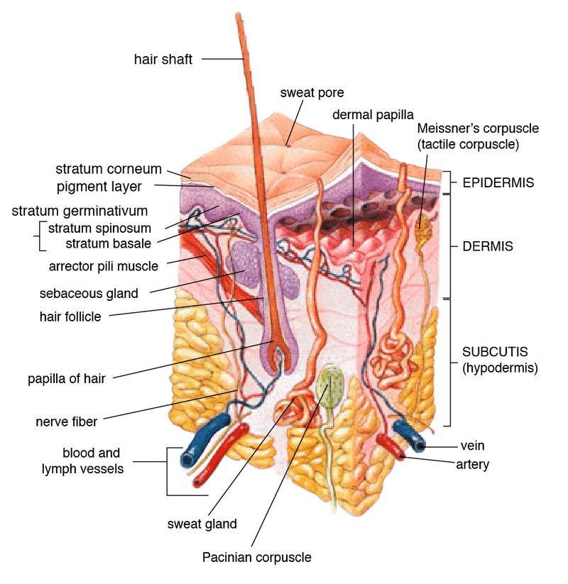
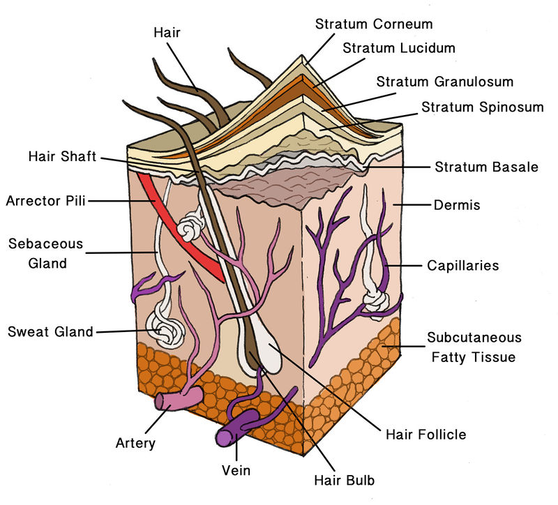
› game › 1566d27ff4Skin Labeling Quiz - PurposeGames.com Oct 07, 2009 · This is an online quiz called Skin Labeling. There is a printable worksheet available for download here so you can take the quiz with pen and paper. From the quiz author. Skin Anatomy: The Layers of Skin and Their Functions - Verywell Health Subcutaneous tissue is the innermost layer of the skin. It is mostly made up of fat, connective tissues, larger blood vessels, and nerves. 5 The majority of your body fat is stored in the subcutaneous layer. It not only insulates you against changing temperatures but protects your muscles and internal organs from impacts and falls. Skin: Cells, layers and histological features | Kenhub The epidermis is the uppermost layer of the skin. Going from deep to superficial, it consists of five layers; basal layer (stratum basale/germinativum) prickle cell layer (stratum spinosum) granular layer (stratum granulosum) clear layer (stratum lucidum) cornified layer (stratum corneum) To remember these layers, check out this mnemonics video:
Skin diagram labeling. Parts of the Microscope with Labeling (also Free Printouts) 5. Knobs (fine and coarse) By adjusting the knob, you can adjust the focus of the microscope. The majority of the microscope models today have the knobs mounted on the same part of the device. Image 5: The circled parts of the microscope are the fine and coarse adjustment knobs. Picture Source: bp.blogspot.com. › en › libraryClitoris: Location, structure, diagram | Kenhub Jul 06, 2022 · The clitoris arises as a pair of crura, two erectile structures which attach to the ischiopubic rami.Anteriorly, each crus converges to form the paired corpora cavernosa of the clitoris, which are collectively known as its body and are enclosed in a layer of dense fibrous connective tissue (known as the tunica albuginea). Human Biology Lab Online | Lab 4 Tissues and Skin When viewing the slides, please follow these steps: Start on the lowest magnification 40x Move to the next higher magnification 100x View the tissue at 400x (Make your sketch of the tissue at this mag.) Can you identify any of the cell structures Select the Labels "On" button Use the slider bars on the side and bottom to scan around the slide. Anatomy, Skin (Integument), Epidermis - StatPearls - NCBI Bookshelf Stratum lucidum, 2-3 cell layers, present in thicker skin found in the palms and soles, is a thin clear layer consisting of eleidin which is a transformation product of keratohyalin. Stratum corneum, 20-30 cell layers, is the uppermost layer, made up of keratin and horny scales made up of dead keratinocytes, known as anucleate squamous cells.
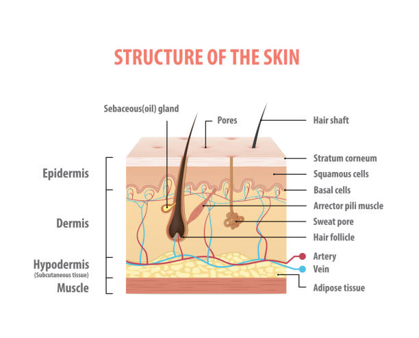
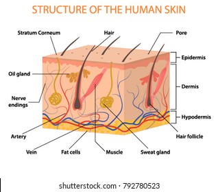
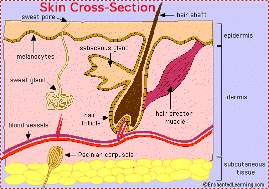
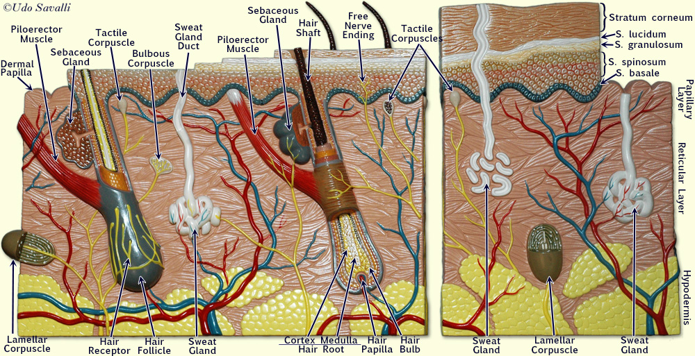
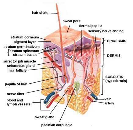
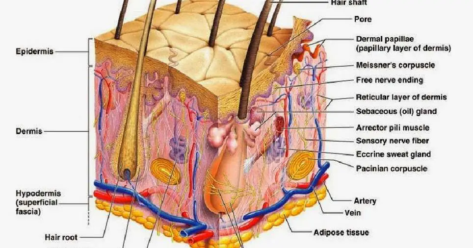
Integumentary system parts: Quizzes and diagrams | Kenhub Labeled diagram of the skin So what's the idea? Spend some time analyzing the skin diagram labeled above. Try to memorize the appearance and location of each structure. Learning the function of each structure will accelerate your ability to memorize, so be sure to check out our detailed article on The Integumentary System parts and functions . Dense Irregular Connective Tissue - AnatomyLearner #1. In the dermis of the thick and thin skin of animals #2. In the fascia, aponeuroses, and joint capsule #3. Fibrous capsules of spleen, testes, ovary, and kidney of animals #4. Lamina propria of the digestive tract of animals. ... If you need more slide images or a labeled diagram of dense connective tissue, ... Anatomy, Skin (Integument) - StatPearls - NCBI Bookshelf The epidermis, the outermost layer of skin, provides a waterproof barrier and contributes to skin tone. The dermis, found beneath the epidermis, contains connective tissue, hair follicles, blood vessels, lymphatic vessels, and sweat glands. The deeper subcutaneous tissue (hypodermis) is made of fat and connective tissue. Structure & Function of Your Skin - American Osteopathic College of ... Skin is a waterproof, flexible, but tough protective covering for your body. Normally the surface is smooth, punctuated only with hair and pores for sweat. A cross-section of skin shows the major parts. It is divided into three layers. The outer layer is the epidermis. The dermis is in the middle and fat forms the innermost layer.
Body Cavities and Membranes: Labeled Diagram, Definitions - EZmed The 3 meningeal layers are labeled with the stars. The outermost layer of the meninges is the dura mater, which is located beneath the skull. Below the dura mater is the arachnoid, which is the middle meningeal layer. There is a space below the arachnoid called the subarachnoid space, and this is where the CSF is located. Vagina Parts | a Diagram and Guide of Female Anatomy - Women's Health 1. The vulva. It's a common misconception that the visible outer parts of the female anatomy is called the vagina. The technical name is actually the vulva. Yours has the job of protecting your ... Microscope Types (with labeled diagrams) and Functions Simple microscope labeled diagram Simple microscope functions It is used in industrial applications like: Watchmakers to assemble watches Cloth industry to count the number of threads or fibers in a cloth Jewelers to examine the finer parts of jewelry Miniature artists to examine and build their work Also used to inspect finer details on products › en › libraryTestes: Anatomy, definition and diagram | Kenhub Aug 22, 2022 · Scrotum is a cutaneous (skin) sac that protects the testes. It consists of two layers: most superficially is the skin, and deeper is the dartos fascia. The dartos fascia contains muscle fibers that contract when it is cold, which results in wrinkling of the scrotal skin and brings the testes closer to the body. The result is a reduction of heat ...
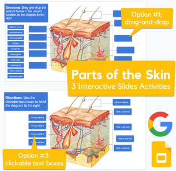
Skin Diagram - drag-and-drop, labeling activity in Slides (advanced)
Foot Pain Diagram - Why Does My Foot Hurt? - Foot-Pain-Explored.com F. Os Trigonum. An Os Trigonum is a small extra bone in the back of the ankle found in 5-10% of people. Causes tenderness and pain behind the ankle particularly when pointing the toes or going up on tiptoes and sometimes swelling. Typically symptomatic in ballet dancers, runners and football players, or after an ankle injury.

Label the Skin Quiz
› 5th-grade-science-games5th grade science games, interactive quiz games 5th grade science games, interactive quiz games, atoms and elements, cells and their functions, ecosystems, electricty and magnetism, extinction and fossils, force and motion

Label the Skin Quiz
› beef-cutsBeef Cuts Chart and Diagram, with Photos, Names, Recipes, and ... Jul 08, 2022 · Ingredients Beef Cuts Chart and Diagram, with Photos, Names, Recipes, and More. Learn all about the most popular beef cuts from our chart, diagram and write up, including popular and alternative names, where the cuts come from on the cow, preferred outdoor cooking methods, their costs relative to each other, and a fantastic recipe for each cut of beef that we’ve taken from around the web.

Skin Structure labeling Diagram | Quizlet
Infographic: Skin cancer body mole map - American Academy of Dermatology When caught early, skin cancer is highly treatable. Download the AAD's body mole map for information on how to check your skin for the signs of skin cancer. Keep track of the spots on your skin and make note of any changes from year-to-year. If you notice a mole that is different from others, or that changes, itches, or bleeds, you should make ...

Skin Diagram with Detailed Illustrations and Clear Labels
› science › biology-diagramFree Biology Diagram Software with Free Templates - EdrawMax Useful templates: There are hundreds of professionally-designed biology diagram templates on EdrawMax. You are likely to find every biology diagram template on EdrawMax. Comprehensive symbol library: An intensive symbol library is available for the biology signs and symbols. You can use the custom color brand palette and all labeling signs in ...

In situ visualization of glucocerebrosidase in human skin ...
Diagram of Human Heart and Blood Circulation in It Four Chambers of the Heart and Blood Circulation. The shape of the human heart is like an upside-down pear, weighing between 7-15 ounces, and is little larger than the size of the fist. It is located between the lungs, in the middle of the chest, behind and slightly to the left of the breast bone. The heart, one of the most significant organs ...

Alat Penghilang Tag/Label Skin Band Micro Untuk Cepat & Efektif di Ngebet Belanja | Tokopedia
The 5 Layers Of Scalp Explained - SkinKraft Of the 5 layers of the scalp [ 1 ], the first three are firmly held together as a unit. The layers of the scalp can be remembered using the mnemonic SCALP: 1. Skin. The skin [ 2] contains several hair follicles and sebaceous glands. A gland is a group of cells that synthesize chemicals needed for bodily functions.
Skin 1: the structure and functions of the skin | Nursing Times
Skin and skin appendage - Knowledge @ AMBOSS The skin is the largest organ of the body, covering an area of approximately 2 m 2.The skin is composed of the cutis (including the dermis and epidermis), subcutaneous tissue, and skin appendages.The epidermis, which is derived from ectoderm, is the outermost layer of the skin and is mainly composed of keratinocytes.The dermis, which is derived from mesoderm, is located underneath the ...
How To Draw Skin Layers | Integumentary System | step by step drawing
› media › 72142Guidance for Industry - Food and Drug Administration section of labeling required by 21 CFR 201.57(c)(3). The guidance provides recommendations ... (e.g., tuberculin skin test before initiating tumor ... flow diagram, or algorithm. The DOSAGE AND ...

ImageQuiz: Labeling Skin Diagram 2016
Skin Under Microscope - The Place to Learn Veterinary Anatomy Online Okay, let's know the microscopic characteristics of the thick and thin skin with the labeled diagrams. Microscopic features of thin skin When you identify the thin skin, you will find the below-mentioned microscopic features. You will find both the epidermis and dermis layers under the microscope while viewing with 10x magnification.

File:Labeled layers of the skin.jpg - Wikimedia Commons
Ask a Dermatologist: How to Read Skincare Ingredient Labels - Byrdie Important Things to Look for on a Label The Order of Ingredients Ingredients are listed from highest to lowest concentration, says Jacqueline Schaffer, an anti-aging expert, best-selling author, and founder of vegan skincare brand Schique. This means if a really great ingredient is listed at the bottom, you're not going to get much benefit from it.

SKIN APPENDAGES HAIR NAILS GLANDS September 23 24
Understanding The Different Layers Of Skin - SkinKraft Skin is the largest organ in your body and acts as a protective barrier. It is a connective tissue that consists of cells, fibres and extracellular matrix [ 1 ]. The three main layers in it are: Epidermis Dermis Hypodermis Functions Of The Skin's Layers 1. Epidermis

21,931 Skin Anatomy Stock Photos, Pictures & Royalty-Free ...
Cosmetics Labeling Regulations | FDA Labeling. This term refers to all labels and other written, printed, or graphic matter on or accompanying a product [FD&C Act, sec. 201 (m); 21 U.S.C. 321 (m)]. Principal Display Panel (PDP). This...
What are the principal layers of the skin? | Socratic
Anatomy of the Epidermis with Pictures - Verywell Health The epidermis is composed of layers of skin cells called keratinocytes. Your skin has four layers of skin cells in the epidermis and an additional fifth layer in areas of thick skin. The four layers of cells, beginning at the bottom, are the stratum basale, stratum spinosum, stratum granulosum, and stratum corneum.
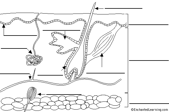
Label Skin Diagram Printout - EnchantedLearning.com
Summary of Cosmetics Labeling Requirements | FDA Off-package ingredient labeling is permitted if the cosmetic is held in tightly compartmented trays or racks, it is not enclosed in a folding carton, and the package surface area is less than 12...
4,031 Skin Diagram Stock Photos, Pictures & Royalty-Free ...
Anatomical Position and Directional Terms - EZmed We will first review the anatomical position, its definition, and look at example labeled diagrams. We will then walk through the different anatomical directional terms used to describe location and movement. We will provide you with labeled diagrams, example body parts, and tricks to learn the directional terms listed below! Medial vs Lateral
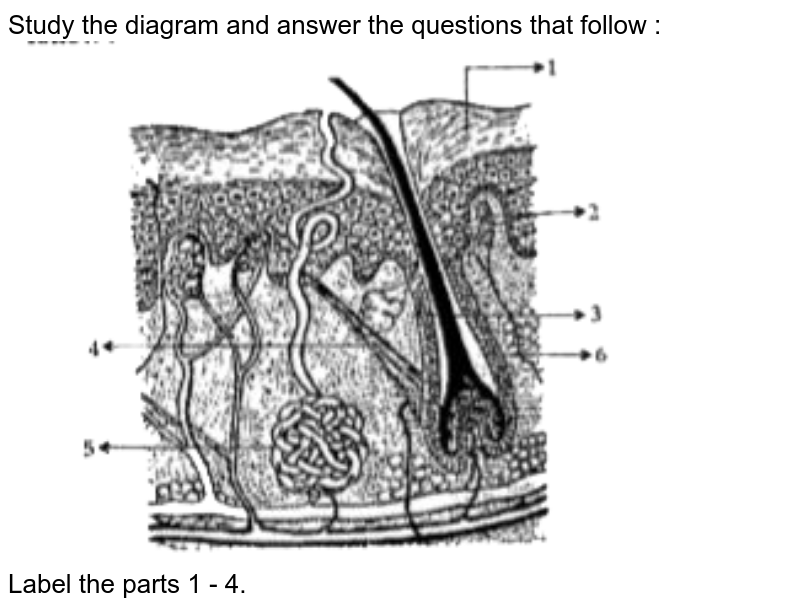
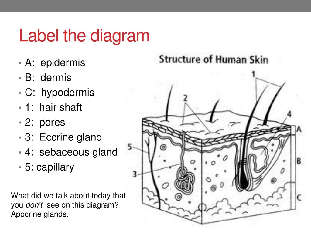
Study the diagram and answer the questions that follow ...
The Skin: 7 Most Important Layers, Functions & Thickness - MedicineNet There are seven layers of skin and each layer serves different functions. The skin is the largest organ in the body and it covers the body's entire external surface. It is made up of seven layers ( [starting from the top layer down to the bottom [deepest] layer): Stratum corneum Stratum lucidum Stratum granulosum Stratum spinosum Stratum basale

label the diagram - Biology - TopperLearning.com | we1gw8ss
WHMIS 2015 - Pictograms : OSH Answers - Canadian Centre for ... Most pictograms have a distinctive red "square set on one of its points" border. Inside this border is a symbol that represents the potential hazard (e.g., fire, health hazard, corrosive, etc.). Together, the symbol and the border are referred to as a pictogram. Pictograms are assigned to specific hazard classes or categories.

Illustration Anatomy Skin Label On White Stock Vector ...
Skin: Cells, layers and histological features | Kenhub The epidermis is the uppermost layer of the skin. Going from deep to superficial, it consists of five layers; basal layer (stratum basale/germinativum) prickle cell layer (stratum spinosum) granular layer (stratum granulosum) clear layer (stratum lucidum) cornified layer (stratum corneum) To remember these layers, check out this mnemonics video:
Draw and label a mammalian skin.
Skin Anatomy: The Layers of Skin and Their Functions - Verywell Health Subcutaneous tissue is the innermost layer of the skin. It is mostly made up of fat, connective tissues, larger blood vessels, and nerves. 5 The majority of your body fat is stored in the subcutaneous layer. It not only insulates you against changing temperatures but protects your muscles and internal organs from impacts and falls.

Given below is a diagrammatic sketch of the vertical section ...
› game › 1566d27ff4Skin Labeling Quiz - PurposeGames.com Oct 07, 2009 · This is an online quiz called Skin Labeling. There is a printable worksheet available for download here so you can take the quiz with pen and paper. From the quiz author.

Anatomy 2017- Unit 2 Label Parts of Skin Diagram Diagram ...

Cross section of human skin with labels | Skin anatomy, Skin ...
Skin: Image Details - NCI Visuals Online
SOLUTION: Integumentary System Identification Skin Diagram ...

Berkas:Diagram of human skin.jpg - Wikipedia bahasa Indonesia ...
Human Skin Layers Vector Illustration Stock Vector (Royalty ...

Schematic representation of the skin layers and appendage ...
Integumentary System- definition, organs, functions, diseases
Human Biology fig. 1.24 - Layers of the skin - English labels ...

Science Is Art
Skin Anatomy - EnchantedLearning.com
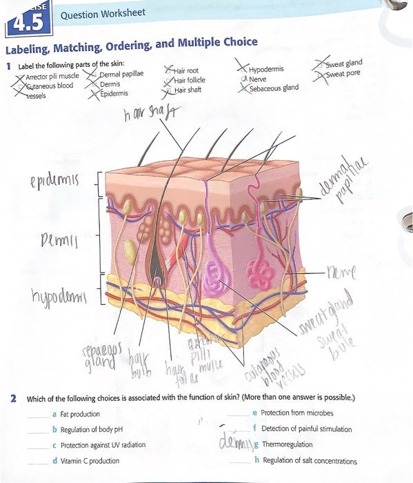
Solved 4.5 Label the following parts of the skin ***Which of ...
BIO201-Skin
Integumentary system review key
PPT - Skin Appendages: Hair, Nails, Glands PowerPoint ...
Skin diagram labeled
Solved] Worksheet The Integumentary System 1. Label the ...

Human skin - Wikipedia

Download Skin Anatomy Cross Section With Labels On White ...

skin labeling Diagram | Quizlet

Epidermis Vector Art Stock Images | Depositphotos
Post a Comment for "42 skin diagram labeling"