39 photomicrograph of thick skin
Solved The photomicrograph of thick skin | Chegg.com Question: The photomicrograph of thick skin · This problem has been solved! · Expert Answer. Who are the experts?Experts are tested by Chegg as specialists in ... Label The Photomicrograph Of Thick Skin. - Stratified squamous ... Label The Photomicrograph Of Thick Skin. - Stratified squamous keratinized epithelium Dehydrated birds will show darker and thinner looking legs, they will feel lighter and the skin will not move freely over the keel. 07.05.2014 · label this group a.
Pathology Outlines - Endometrial hyperplasia Feb 20, 2020 · Thick walled blood vessels Endometrial polyps can contain foci of AH / EIN Disordered proliferative endometrium: No well delineated criteria Histologically considered as degree below hyperplasia without atypia on a shared morphologic spectrum and distinction is often not reproducible Both have similar treatment (exogenous progestin)

Photomicrograph of thick skin
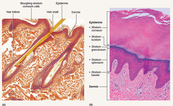
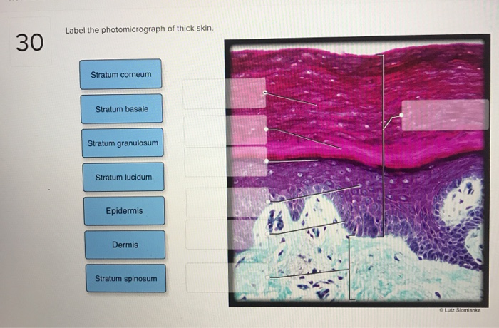
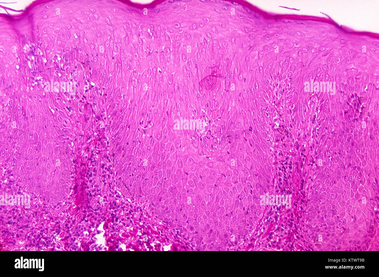
photomicrograph of the epidermal layer in thick skin photomicrograph of the epidermal layer in thick skin Diagram | Quizlet photomicrograph of the epidermal layer in thick skin STUDY Learn Write Test PLAY Match + − Created by abba_dabba_17 Terms in this set (6) stratum corneum ... stratum lucidum ... stratum granulosum ... stratum spinosum ... stratum basale ... dermis ... OTHER SETS BY THIS CREATOR Skin overview 4 | Digital Histology A diagrammatic representation of thin skin and a photomicrograph of a H&E stained section illustrate the reduced thickness of the strata in thin skin and the absence of stratum lucidum as a distinct layer. 400x - Stratum spinosum Skin can be classified as either thick or thin, depending on the thickness of the epidermal layer. Solved Label the photomicrograph of thick skin. Stratum Question: Label the photomicrograph of thick skin. Stratum corneum Stratum basale Stratum granulosum Stratum lucidum Epidermis Dermis Stratum spinosum.
Photomicrograph of thick skin. Elastofibroma dorsi - Wikipedia Elastofibroma dorsi is an ill-defined fibroelastic tumor-like condition made up of enlarged and irregular elastic fibers. The World Health Organization, 2020, has classified elastofibroma tumors as one specific type of the fibroblastic and myofibroblastic tumors. Label The Photomicrograph Of Thick Skin / Solved Label The ... - Blogger The outer layer of cells in this micrograph is the thinnest layer and. Thick skin is found only on the palms of the hands and the soles of the feet. 1 answer to label the photomicrograph of thin skin. Label the photomicrograph of thick skin. The stratum lucidum (only found in thick skin), and the stratum corneum. Solved 21. Label the photomicrograph of thick skin - Chegg Question: 21. Label the photomicrograph of thick skin, Epidermis Stratum basale Stratum granulosum Stratum corneum Suatum spinosum to Dermis Stratum lucidum ... Label The Photomicrograph - Mr. Hill's Biology Blog: Our cells "inner skin" Label the photomicrograph of thick skin. Label the structures of the skin and … The disc with the seeds can be attached to a clinostat, as shown below in figure 6.2. Specimens prepared with fixatives that contain 50% ethyl alcohol, eg, saccomanno fixative, are … Place the following layers in order from superficial to deep. 3.1 (a) (i) it is ...
SKIN | The Big Picture: Histology | AccessBiomedical Science | McGraw ... A subcutaneous layer of loose connective tissue below the dermis that attaches the skin to underlying tissues. Skin contains various appendages derived from epidermis, including sweat glands, hair follicles and sebaceous glands. Skin is classified as either thick or thin. Thick skin is found on the palms and the soles. In the photomicrograph of a portion of thick skin - Course Hero Section Reference 1: Sec 5.1 Structure of the Skin. 33) In the photomicrograph of a portion of thick skin shown below, which layer is the stratum basale? a) Ab) B c) D d) Ee) F Answer: d. Difficulty: Medium Study Objective 1: SO 5.1 Describe the general structure of the skin. Sebaceous Gland Label The Photomicrograph Of Thin Skin - Integumentary ... Name the 4 layers of thin skin in both the cartoon and the photomicrograph. Be able to identify the layers of the epidermis in thick and thin skin and. Long thin myoepithelial cells are arranged helically around the periphery between the . Dermis duct of sebaceous gland hair follicle sebaceous gland hair epidermis. This problem has been solved! Label The Photomicrograph Of Thick Skin - Faktor yang The epidermis of thick skin has five layers: Take several photomicrographs of thin skin at this magnification. Stratum basale, stratum spinosum, stratum granulosum, stratum lucidum, and stratum . Layers of skin and label the. Thick skin · stratum basale (also known as s.
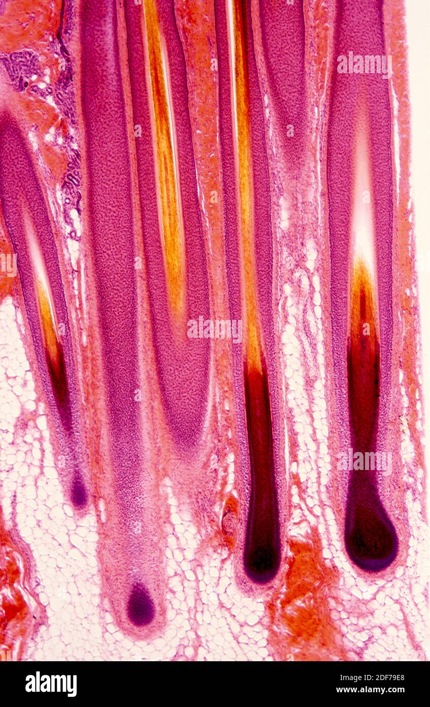
(PDF) Paniker's Textbook of Medical Parasitology, 7th Edition ... Paniker's Textbook of Medical Parasitology, 7th Edition (2013) [PDF] Label The Photomicrograph Of Thick Skin. - Blogger 1 answer to label the photomicrograph of thin skin. The epidermis, made of closely packed epithelial cells, and the dermis, made of dense, irregular connective tissue . Epidermis Of Thick Skin from eugraph.com The skin is composed of two main layers: Thick skin showing epithelial detail. Practice labeling the layers of the skin. Anime Windows 10 11 Themes Page 2 Themepack Me - Blogger A free desktop customization program for wi. Learn how to make your windows desktop look way more interesting and beautiful with this anime setup. See more ideas about anime, windows themes, online themes. Share anime theme for windows 7 and windows 8 and anime aimp3 skin. Here's 36 best free windows 10 anime theme free download 2022 · 1. Block1/Fig 10. Dermis of thick skin - Kaohsiung Medical University Fig 10. Dermis of thick skin. This photomicrograph showsthe connective tissue of the skin, referred to as dermis,stained to show the nature and distribution of the elasticfibers (EF), which appear purple. The collagen fibers (CF)have been stained by eosin, and the two fiber types are easilydifferentiated. The elastic fibers of the dermis have a 3Dinterlacing configuration, thus the variety of ...
Thick Skin - Labeled - Histology | Thick skin, Epidermis ...
American Journal of Veterinary Research | AVMA This month's cover image is a photomicrograph of a skin sample obtained from the incision site of a koi 2 weeks after coelioscopy. The image depicts poor healing; the epithelium is incomplete, and the wound surface is partially covered by a layer of serofibrinous crust and cellular debris.

Pigmentation in plexiform neurofibroma following Blaschko's ...
Solved Label the photomicrograph of thick skin. Stratum Question: Label the photomicrograph of thick skin. Stratum spinosum Epidermis Dermis Stratum corneum Stratum granulosum Stratum basale Stratum lucidum · This ...

Epidermal abnormalities after in utero TCDD exposure. (A ...
(Get Answer) - Label The Photomicrograph Of Thick Skin. Stratum ... Procedure Microscopy of Thick Skin obtain a prepared slide of thick skin (which may be labeled "Palmar Skin"), and examine it with the naked eye to get oriented. Once you are oriented, place the slide on the stage of the microscope, and scan it on... Posted 7 months ago Recent Questions in Computer Graphics and Multimedia Applications Q:

photomicrograph of thick skin Diagram | Quizlet
Solved Label the photomicrograph of thick skin. Epidermis | Chegg.com Expert Answer. Answer - There are two types of skin in human body : Thick skin Thin skin …. View the full answer. Transcribed image text: Label the photomicrograph of thick skin. Epidermis Stratum Basale Stratum lucidum Stratum cometim Stratum spinosu Stratus grinulosum Stratum corneum 10 Stratum lucidum Stratum granulosum Stratum spinosum ...

a): A photomicrograph of a vertical section at the skin of upper ...
Palmar Skin - Florida State University Indeed, the skin that lines the palms of a human is typically 0.8 to 1.4 millimeters thick, while most other parts of the body are only protected by an integument 0.1 millimeters thick. Within palmar skin, five morphologically discrete layers of tissue exist. The outermost layer is the stratum corneum, which is predominantly comprised of dead ...
Solved 94 Exercise ? The hair shaft and the hair root have ...
Anatomy, Skin (Integument), Epidermis - StatPearls - NCBI Bookshelf The thickness of each layer of the skin varies depending on body region and categorized based on the thickness of the epidermal and dermal layers. Hairless skin found in the palms of the hands and soles of the feet is thickest because the epidermis contains an extra layer, the stratum lucidum.
Pharmaceutics | Free Full-Text | Plasminogen-Loaded Fibrin ...
Chagas disease - Wikipedia Benznidazole and nifurtimox often cause side effects, including skin disorders, digestive system irritation, and neurological symptoms, which can result in treatment being discontinued. [2] [8] As of 2019 [update] , new drugs for Chagas disease are under development, and experimental vaccines have been studied in animal models.

A) A photomicrograph of a control thin skin of adult albino ...
In the photomicrograph shown below which layer is - Course Hero Collagen is the strongest fiber in the skin. 2. Blood vessels in the dermis supply the epidermis with nutrients. 3. The arrangement of epidermal ridges, mixed with sweat create fingerprints. 4. The papillary layer is the strongest portion of the dermis. 5. The reticular layer promotes stretching of the skin.
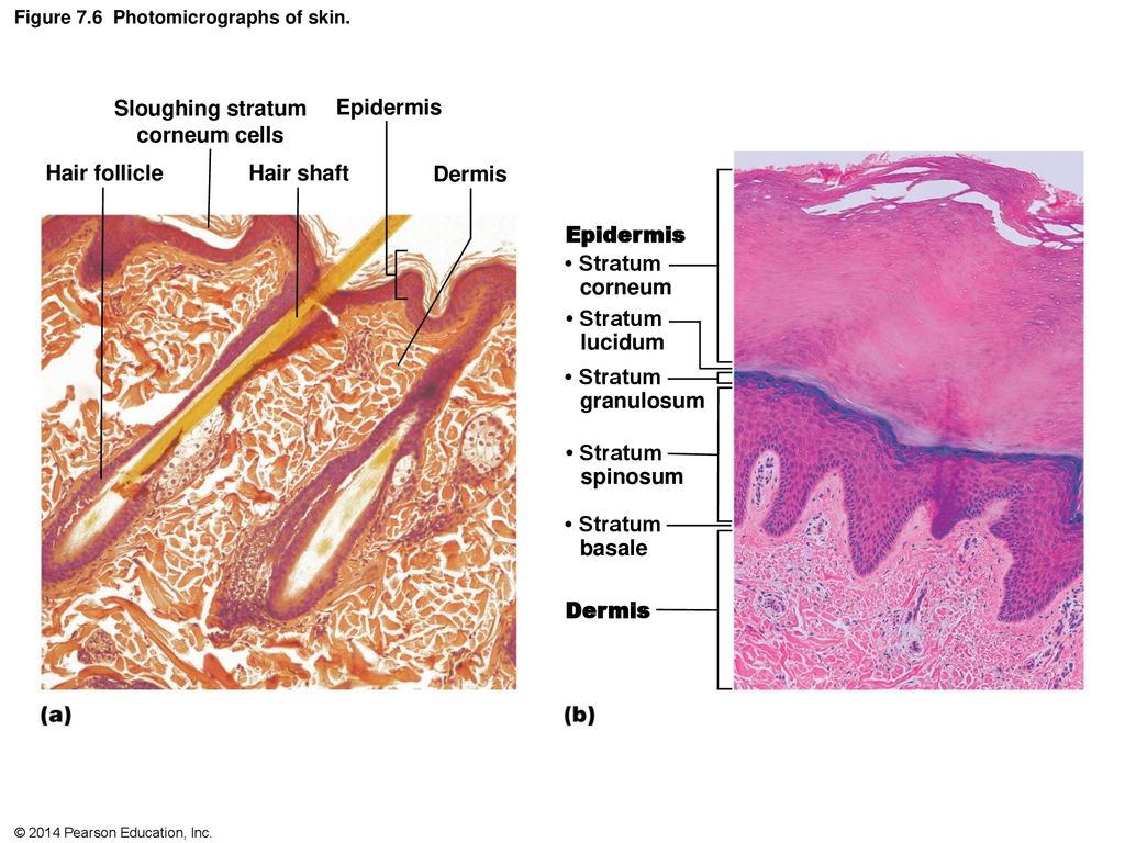
Solved Figure 7.6: (a) Thin skin with hairs (120X). (b ...
Solved Label the photomicrograph of thick skin. Stratum - Chegg Question: Label the photomicrograph of thick skin. Stratum granulosum Stratum spinosum Epidermis Stratum corneum Stratum basale Dermis Stratum lucidum ...

4,731 Photomicrograph Photos - Free & Royalty-Free Stock ...
Skin overview 3 | Digital Histology This image compares a diagrammatic representation of thick skin with a photomicrograph of a hematoxylin and eosin-stained section of primate skin. 200x - Papillary layer of dermis Skin can be classified as either thick or thin, depending on the thickness of the epidermal layer.
Hair follicle micrograph hi-res stock photography and images ...
Photomicrograph of Thick Skin - Printable - PurposeGames.com This is a printable worksheet made from a PurposeGames Quiz. To play the game online, visit Photomicrograph of Thick Skin Download Printable Worksheet Please note! You can modify the printable worksheet to your liking before downloading. Download Worksheet Include correct answers on separate page About this Worksheet

Photomicrographs of the skin wound area on the 10th day. a,b ...
The differences between thick and thin skin - University of Leeds Dermis: Thick skin has a thinner dermis than thin skin, and does not contain hairs, sebaceous glands, or apocrine sweat glands. Thick skin is only found in areas where there is a lot of abrasion - fingertips, palms and the soles of your feet. show labels This is a picture of an H&E stained section of the epidermis of thin skin.
Solved Label the photomicrograph of thick skin. Stratum ...
Block1/Fig 11. Hypodermis of the thick skin. - Kaohsiung Medical University Fig 11. Hypodermis of the thick skin. The lower magnification photomicrograph shows part of the hypodermisof the thick skin. It contains abundant adipocytes. Theadipocyte (Ad) nucleus is compressed and displaced to oneside of the stored lipid droplets and the cytoplasm includingorganelles is reduced to a small rim (Fig 11c). Fig 11ashows several adipocytes and nerve fiber bundles (NB).Fig 11b ...
Nanomaterials | Free Full-Text | Development of Epidermal ...
Photomicrograph of Thick Skin Quiz - PurposeGames.com This is an online quiz called Photomicrograph of Thick Skin There is a printable worksheet available for download here so you can take the quiz with pen and paper. Your Skills & Rank Total Points 0 Get started! Today's Rank -- 0 Today 's Points One of us! Game Points 6 You need to get 100% to score the 6 points available Actions Add to Playlist

Chapter 5: Structure of skin (5.1) and functions of skin (5.4 ...
Layers of the Skin | Anatomy and Physiology I - Lumen Learning Skin that has four layers of cells is referred to as "thin skin.". From deep to superficial, these layers are the stratum basale, stratum spinosum, stratum granulosum, and stratum corneum. Most of the skin can be classified as thin skin. "Thick skin" is found only on the palms of the hands and the soles of the feet.

Block1/Fig 10. Dermis of thick skin
Pathologic features of diabetic thick skin - ScienceDirect Unlike scleroderma, diabetic thick skin resulted in small fiber sizes (<60 nm) only rarely, and bimodality of fiber sizes was not seen. Our study shows that diabetic thick skin is a specific eritity with characteristic light microscopic and ultrastructural features that are totally different from those seen in Scleroderma.
![Solved] In the Photomicrograph of a Portion of Thick Skin ...](https://d2lvgg3v3hfg70.cloudfront.net/TB6088/11ea5de3_9811_9602_9534_8b82ce0ec6c4_TB6088_00.jpg)
Solved] In the Photomicrograph of a Portion of Thick Skin ...
Mange in the Red Fox | Wildlife Online Summary: There are several different forms of mange, each caused by a different species of mite, but sarcoptic mange most commonly affects foxes. Sarcoptic mange is a skin disease caused by the small (2 to 4 mm, or less than one-quarter of an inch) parasitic mite Sarcoptes scabiei, several thousand of which may burrow into a single square-centimetre of skin.

PHOTOMICROGRAPH - Definition and synonyms of photomicrograph ...
Solved Label the photomicrograph of thick skin | Chegg.com Expert Answer. 91% (11 ratings) Transcribed image text: Label the photomicrograph of thick skin.
Animals | Free Full-Text | Morphology and Histology of the ...
photomicrograph of thick skin Diagram | Quizlet photomicrograph of thick skin Diagram | Quizlet photomicrograph of thick skin STUDY Learn Write Test PLAY Match Created by mckennawebber Terms in this set (7) epidermis (stratum corneum - stratum basale) ... stratum corneum ... stratum lucidum ... stratum granulosum ... stratum spinosum ... stratum basale ... dermis ... njordan6 mckennawebber

Thin Skin. Hematoxylin-eosin Stock Photo - Image of ...
Human Skin Microscope Pictures, Images and Stock Photos Low magnification micrograph of a human thick skin stained with the Cajal-Gallego's method. The epidermis (with a thick stratum corneum) and the dense irregular connective tissue of the dermis are clearly seen ... Melanoma of a human Melanoma of a human, photomicrograph panorama as seen under the microscope, 200x zoom. human skin microscope ...

photomicrograph of the epidermal layer in thick skin Diagram ...
Solved Label the photomicrograph of thick skin. Stratum Question: Label the photomicrograph of thick skin. Stratum corneum Stratum basale Stratum granulosum Stratum lucidum Epidermis Dermis Stratum spinosum.

EXERCISE 4 QUIZ Flashcards | Quizlet
Skin overview 4 | Digital Histology A diagrammatic representation of thin skin and a photomicrograph of a H&E stained section illustrate the reduced thickness of the strata in thin skin and the absence of stratum lucidum as a distinct layer. 400x - Stratum spinosum Skin can be classified as either thick or thin, depending on the thickness of the epidermal layer.
Histology of major organ systems of Nothobranchius fishes ...
photomicrograph of the epidermal layer in thick skin photomicrograph of the epidermal layer in thick skin Diagram | Quizlet photomicrograph of the epidermal layer in thick skin STUDY Learn Write Test PLAY Match + − Created by abba_dabba_17 Terms in this set (6) stratum corneum ... stratum lucidum ... stratum granulosum ... stratum spinosum ... stratum basale ... dermis ... OTHER SETS BY THIS CREATOR
Sebaceous (oil) gland • Hair follicle - ppt download

Photomicrograph of histological section of the wounds on day ...

Promotion of skin regeneration in diabetic rats by ...
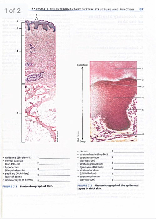
Solved EXERCISE 7 THE INTEGUMENTARY SYSTEM STRUCTURE AND ...
Scrotal leiomyoma: a rare cause of scrotal swelling | African ...

Photomicrographs of skin section obtained from patient with ...
The Integumentary System | SpringerLink

Photomicrographs of interfollicular control rabbit skin at 0 ...

On A, photomicrograph of cross section of the apex of the ...

Skin Epidermis Dermis Subcutaneous Layer Thick Skin 25x At ...

Photomicrographs of the healing wounds on days 14 and 28 ...

Chapter 13, Page 8 - HistologyOLM

Intraosseous epidermoid cysts of adjacent digits in a dog ...
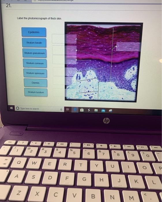
Solved 21. Label the photomicrograph of thick skin, | Chegg.com

Pin by nico x. on Anatomy | Games, Tetris, Anatomy
Skin histology hi-res stock photography and images - Page 2 ...
Post a Comment for "39 photomicrograph of thick skin"