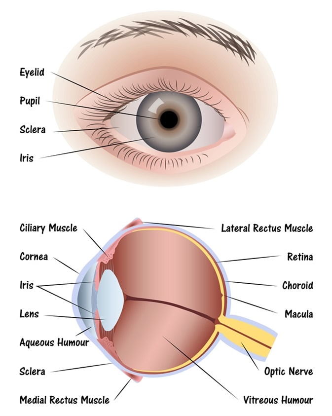
43 easy diagram of human eye with labelling
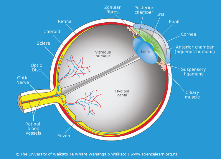
eye diagram with labelling senses special anatomy vision chapter system diagram exercise visual flashcards notecards easy easynotecards. Orientation And Mobility Family Booklet ... eye diagram blank human labeled anatomy label eyeball worksheet quiz labels drawing purposegames answers printable parts ear activities eyes coloring. Eyes — Kidcyber ... Labelling the eye — Science Learning Hub Labelling the eye. Use this interactive to label different parts of the human eye. Drag and drop the text labels onto the boxes next to the diagram. Selecting or hovering over a box will highlight each area in the diagram. The human eye has several structures that enable entering light energy to be converted to electrochemical energy.
How to Draw Human Eye Diagram Easy Step - YouTube Thanks for watching our Channel. human eye diagram,human eye diagram for class 8,construction of human eye diagram,drawing human eye drawing easy step,human ...

Easy diagram of human eye with labelling
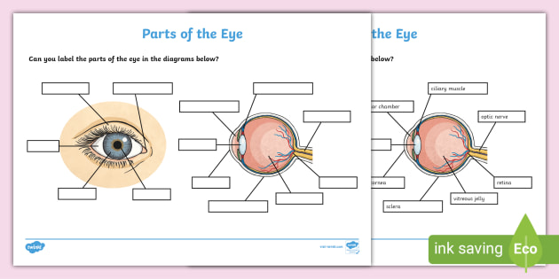
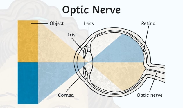
Label the Eye Diagram - Enchanted Learning Label the Eye Diagram. Human Anatomy. Read the definitions, then label the eye anatomy diagram below. Cornea - the clear, dome-shaped tissue covering the front of the eye. Iris - the colored part of the eye - it controls the amount of light that enters the eye by changing the size of the pupil. Lens - a crystalline structure located just behind ... FREE! - The Human Eye Labelling Activity - Twinkl In this resource, you'll find a 2-page PDF that is easy to download, print out, and use immediately with your class. The first page is a labelling exercise with two diagrams of the human eye. One is a view from the outside, and the other is a more detailed cross-section. Challenge learners to label the parts of the eye diagram. How to Draw Human Eye Diagram Step by step for beginners 2. Eye bands Ray of light to form sharp image. 3. Eye sent the information about the image to the brain. Different parts of Eyes :- 1. Cornea - a transparent protective membrane is called cornea....
Easy diagram of human eye with labelling. Diagram of the Eye - Home - Lions Eye Institute Instructions. Click the parts of the eye to see a description for each. Hover the diagram to zoom. Iris. The iris is the coloured part of the eye which surrounds the pupil. It controls light levels inside the eye, similar to the aperture on a camera. The iris contains tiny muscles that widen and narrow the pupil size. The Eye - diagram to label | Teaching Resources File previews. pdf, 2.94 MB. Diagram of eye with key words to use in labelling it. Tes classic free licence. Pin on Five Senses - Pinterest Eye Diagram - Differentiated Worksheets and EASEL Activities Description Use these simple eye diagrams to help students learn about the human eye. Three differentiated worksheets are included: 1. Write the words using a word bank 2. Cut and paste the words 3. Quiz: Label The Parts Of The Eye - ProProfs How much did you get to understand about the human eye? Take up this quiz and find out! Questions and Answers. 1. A is pointing to what part of the eye? A. Cornea. B. Optic Nerve.
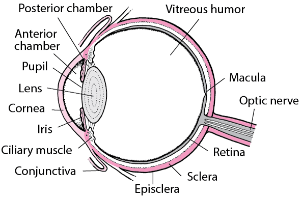
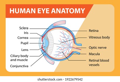
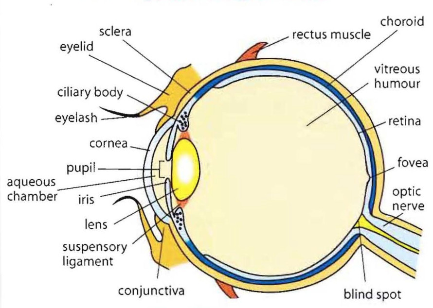
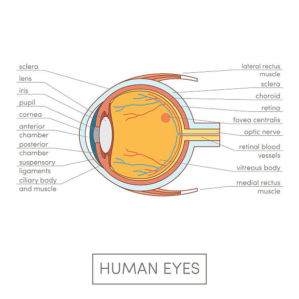
Human Eye: Structure of Human Eye (With Diagram) | Biology The human eye is a very sensitive and delicate organ suspended in the eye socket which protects it from injuries. It essentially consists of CORNEA, LENS & RETINA besides many other parts such as Iris, Pupil and aqueous humour, vituous humour etc. Each one has got a specific function. A section of the eye is as shown in Fig. 2.2. Structure and Functions of Human Eye with labelled Diagram Human Eye Diagram: Contrary to popular belief, the eyes are not perfectly spherical; instead, it is made up of two separate segments fused together.Explore: Facts About The Eye. To understand more in detail about our eye and how our eye functions, we need to look into the structure of the human eye. Recommended Video: File:Simple diagram of human eye multilingual.svg - Wikimedia Size of this PNG preview of this SVG file: 370 × 429 pixels. Other resolutions: 207 × 240 pixels | 414 × 480 pixels | 662 × 768 pixels | 883 × 1,024 pixels | 1,766 × 2,048 pixels. Original file (SVG file, nominally 370 × 429 pixels, file size: 1.14 MB) File information. Structured data. How to Draw Human Eyes: 9 Steps (with Pictures) - wikiHow 1. Draw the upper line of the eye first. Draw an arc shape, as shown in the image. 2. Draw the lower line of the eye. Use the same arc drawing technique but instead this time do it upside down. 3. Draw the inside of the eye (the iris). Draw a circle, lightly, inside what you have just finished drawing.
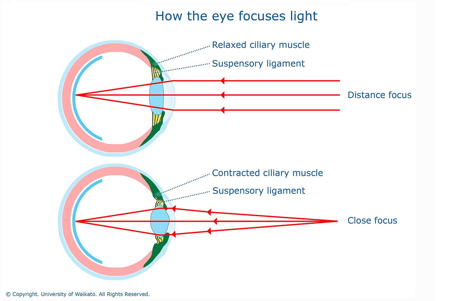
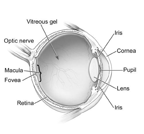
Human Ear Diagram - Bodytomy The Structure of Human Ear. Helix: It is the prominent outer rim of the external ear. Antihelix: It is the cartilage curve that is situated parallel to the helix. Crus of the Helix: It is the landmark of the outer ear, situated right above the pointy protrusion known as the tragus. Auditory Ossicles: The three small bones in the middle ear ... Draw a labelled diagram of human eye and explain the image ... - Toppr Ask the figure below shows the labelled diagram of the human eye . the main parts of eye are :-. Cornea , Iris , Pupil , Ciliary muscles , Eye lens , retina and optical nerves which are labelled in the diagram below . Image Formation : The light rays coming from object enter through of eye , pass through the pupil of the eye and fall on the eye ... Human Eye Anatomy Pictures, Images and Stock Photos Browse 8,092 human eye anatomy stock photos and images available, or search for vision or retina to find more great stock photos and pictures. Anatomy of human eye and descriptions. Components of human eye. Illustration about Anatomy and Physiology. Parts of the eye, labeled vector illustration diagram. PDF Parts of the Eye - National Eye Institute | National Eye Institute Eye Diagram Handout Author: National Eye Health Education Program of the National Eye Institute, National Institutes of Health Subject: Handout illustrating parts of the eye Keywords: parts of the eye, eye diagram, vitreous gel, iris, cornea, pupil, lens, optic nerve, macula, retina Created Date: 12/16/2011 12:39:09 PM
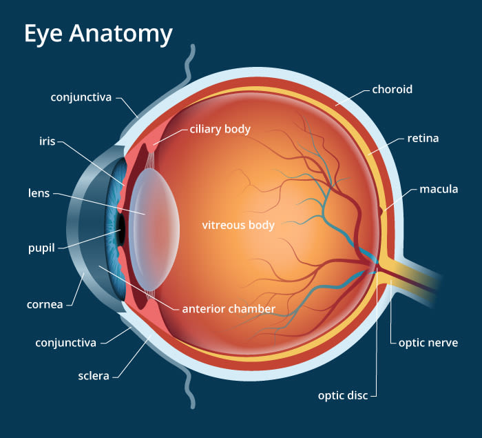
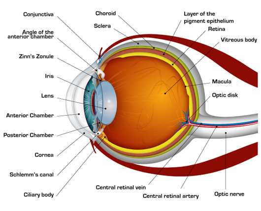
Eye Anatomy: 16 Parts of the Eye & Their Functions The following are parts of the human eyes and their functions: 1. Conjunctiva The conjunctiva is the membrane covering the sclera (white portion of your eye). The conjunctiva also covers the interior of your eyelids. Conjunctivitis, often known as pink eye, occurs when this thin membrane becomes inflamed or swollen.
Eye Diagram Quiz - ProProfs Try this amazing Eye Diagram Quiz quiz which has been attempted 5004 times by avid quiz takers. ... Label The Parts Of The Eye. People say that the eyes are the windows to a person's soul. ... How much did you get to understand about the human eye?... Questions: 8 | Attempts: 44770 | Last updated: Mar 22, 2022 . Sample Question. A is pointing ...
Eye Diagram Teaching Resources | Teachers Pay Teachers Anatomy of the Eye Diagrams for Coloring/Labeling, with Reference and Summary by Homemade For Play 8 $1.95 PDF This printable contains 13 clear and simple cross sectional diagrams of the human eye. They photocopy well and are great for use as a labeling and coloring exercise for your students.
PDF Eye Anatomy Handout - National Eye Institute of light entering the eye. Lens: The lens is a clear part of the eye behind the iris that helps to focus light, or an image, on the retina. Macula: The macula is the small, sensitive area of the retina that gives central vision. It is located in the center of the retina. Optic nerve: The optic nerve is the largest sensory nerve of the eye.
6,782 Human eye diagram Images, Stock Photos & Vectors - Shutterstock Human eye diagram royalty-free images 6,782 human eye diagram stock photos, vectors, and illustrations are available royalty-free. See human eye diagram stock video clips Image type Orientation People Artists More Sort by Biology Healthcare and Medical Icons and Graphics Diseases, Viruses, and Disorders human eye anatomy eye 3d rendering retina
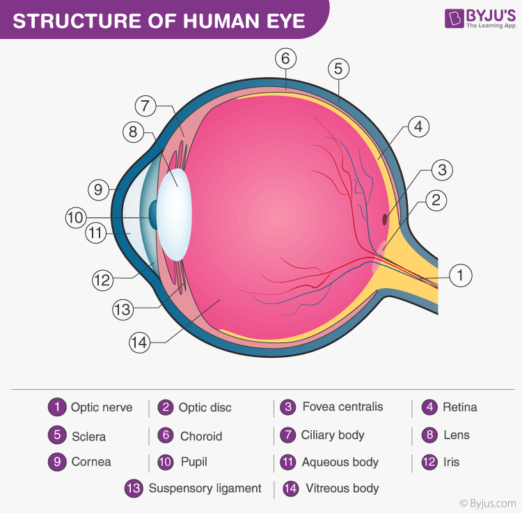
Eye Diagram With Labels and detailed description - BYJUS A brief description of the eye along with a well-labelled diagram is given below for reference. Well-Labelled Diagram of Eye The anterior chamber of the eye is the space between the cornea and the iris and is filled with a lubricating fluid, aqueous humour. The vascular layer of the eye, known as the choroid contains the connective tissue.
The Human Eye (Eyeball) Diagram, Parts and Pictures Diagram of the orbit (eyeball socket) in the skull The orbit is made up of several bones of the skull : Frontal bone at the top (superiorly) Zygomatic bone on the front and outer side (anterolaterally) Maxilla on the front and inner side (anteromedially) Lacrimal and nasal bones on the back and inner side (posteromedially)
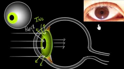
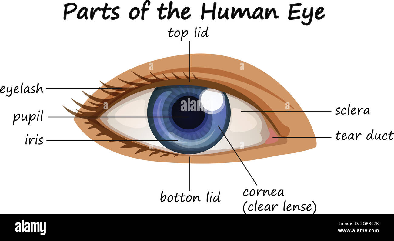
The Eyes (Human Anatomy): Diagram, Optic Nerve, Iris, Cornea ... - WebMD The front part (what you see in the mirror) includes: Iris: the colored part. Cornea: a clear dome over the iris. Pupil: the black circular opening in the iris that lets light in. Sclera: the ...
Labeled Eye Diagram | Science Trends The human eye is composed of many different parts that work together to interpret the world around us. What you want to interpret as a major part of the human eye is somewhat up to the individual, but in general there are seven parts of the human eye: the cornea, the pupil, the iris, the lens, the vitreous humor, the retina, and the sclera. Let's take a closer look at each of these ...
Labelling the eye — Science Learning Hub Labelling the eye Add to collection The human eye contains structures that allow it to perceive light, movement and colour differences. In this activity, students use online or paper resources to identity and label the main parts of the human eye. By the end of this activity, students should be able to: identify the main parts of the human eye
How to Draw Human Eye Diagram Step by step for beginners 2. Eye bands Ray of light to form sharp image. 3. Eye sent the information about the image to the brain. Different parts of Eyes :- 1. Cornea - a transparent protective membrane is called cornea....
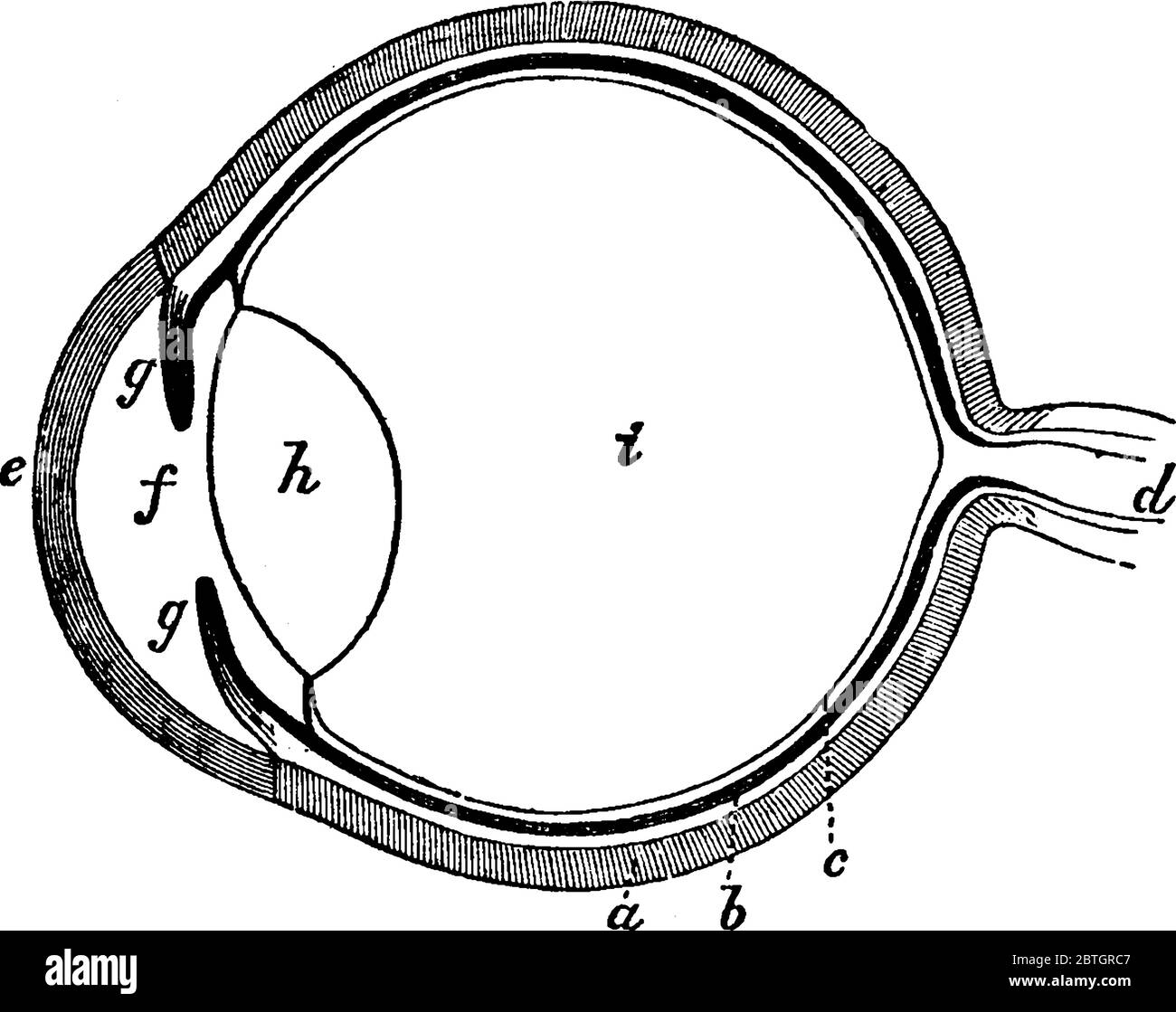
FREE! - The Human Eye Labelling Activity - Twinkl In this resource, you'll find a 2-page PDF that is easy to download, print out, and use immediately with your class. The first page is a labelling exercise with two diagrams of the human eye. One is a view from the outside, and the other is a more detailed cross-section. Challenge learners to label the parts of the eye diagram.
Label the Eye Diagram - Enchanted Learning Label the Eye Diagram. Human Anatomy. Read the definitions, then label the eye anatomy diagram below. Cornea - the clear, dome-shaped tissue covering the front of the eye. Iris - the colored part of the eye - it controls the amount of light that enters the eye by changing the size of the pupil. Lens - a crystalline structure located just behind ...










/GettyImages-695204442-b9320f82932c49bcac765167b95f4af6.jpg)

![Cross sectional diagram of human eye [1]. | Download ...](https://www.researchgate.net/publication/276541864/figure/fig1/AS:612895498964992@1523137082339/Cross-sectional-diagram-of-human-eye-1.png)



Post a Comment for "43 easy diagram of human eye with labelling"