42 labeled gel electrophoresis photo
gel electrophoresis | Britannica gel electrophoresis, any of several techniques used to separate molecules of DNA, RNA, or protein on the basis of their size or electric charge. Gel electrophoresis has a variety of applications; for example, it is used in DNA fingerprinting and the detection of genetic variants and proteins involved in health and disease as well as in the detection and purification of nucleic acids and ... Gel Electrophoresis: Basics & Steps | SchoolWorkHelper Aragonese and the buffer are mixed together and microwaved to create the gel. It is poured into a mold and has a "comb" placed in it to make holes for the DNA to be inserted. Once it has cooled the comb is removed. The gel is then placed in the gel electrophoresis box and buffer solution is poured onto it. The buffer conducts the current.
Gel electrophoresis: Types, introduction and their applications Pulsed-field gel electrophoresis (PFGE) This technique was developed by Shwartz and Cantor in 1984. Separation of DNA in agarose gel by altering the strength and direction of the electrical field between electrodes. It is used to separate high molecular weight DNA of several megabases, even whole chromosomes.
Labeled gel electrophoresis photo
3 Ways to Read Gel Electrophoresis Bands - wikiHow Gel electrophoresis is a type of biotechnology that separates molecules based on their size to interpret an organism's DNA. An enzyme is used to separate a strand of DNA from a source and the DNA is suspended in a dye. Then, the dye is applied to a negatively-charged gel on one side of a sheet. Addgene: Protocol - How to Run an Agarose Gel Run the gel at 80-150 V until the dye line is approximately 75-80% of the way down the gel. A typical run time is about 1-1.5 hours, depending on the gel concentration and voltage. Note: Black is negative, red is positive. The DNA is negatively charged and will run towards the positive electrode. Always Run to Red. Lab 7 - Gel Electrophoresis - Biology Lab Notebook - Google Abstract. In this lab, a liquid agarose base was used to create a gel base for an electrophoresis procedure using different strands of DNA. Gel electrophoresis is used to separate macromolecules into fragments based on their size. The DNA samples were placed in the wells of the agarose gel at the negative end, and then had a current run through ...
Labeled gel electrophoresis photo. Gel Electrophoresis Lab Report - Google Docs This lab exposes us to DNA technology. Backround Gel electrophoresis is used to separate macromolecules like DNA or RNA by size or proteins by charge. To examine DNA and RNA, the fragments are... Gel electrophoresis — Science Learning Hub This technique is called SDS-PAGE (SDS-Polyacrylamide gel electrophoresis). Small protein molecules move more quickly through the gel than larger proteins, resulting in a series of 'bands'. Each band contains a protein of a particular size. These can be compared with standards of known sizes. Gel Electrophoresis - an overview | ScienceDirect Topics Gel electrophoresis is an analytical technique that allows size separation of DNA as well as other macromolecules. For gel electrophoresis, a DNA sample is loaded at one end of a gel matrix (usually agarose or acrylamide) that provides a uniform pore size through which the DNA molecules can move. PDF Gel electrophoresis: sort and see the DNA 10. On the gel picture below, (a) circle the smallest fragment produced by a restriction enzyme and label it "smallest." (b) circle the largest fragment produced by a restriction enzyme and label it "largest." 11. In one or two sentences, summarize the technique of gel electrophoresis. Student answers DNA restriction fragment size chart
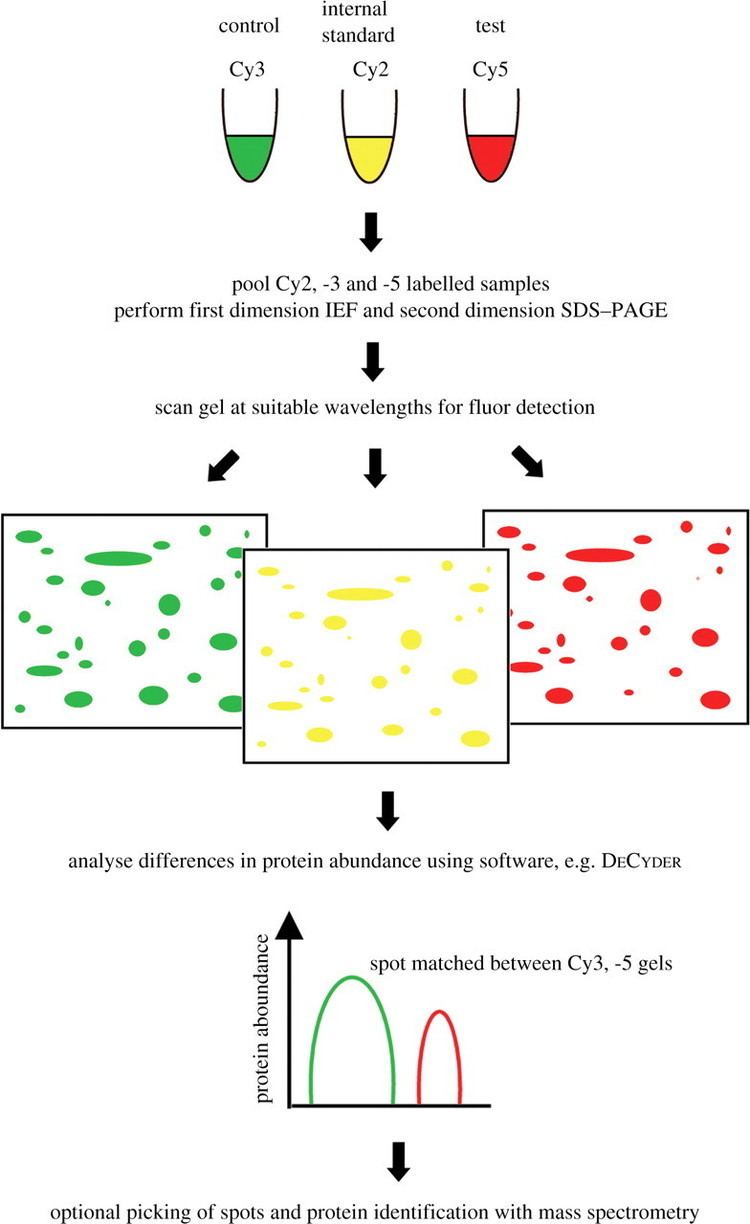
Two-Dimensional Gel Electrophoresis - an overview | ScienceDirect Topics Two-dimensional gel electrophoresis (2DGel) is a successful method used for the detection and analysis of proteins. It has been designed as a combination of the 2DGel, IEF and SDS-PAGE methods, and is used in the analysis of complex protein mixtures. In the first step, protein is separated into its charges with IEF, whereas in the second step ... Gel electrophoresis Images, Stock Photos & Vectors - Shutterstock 768 gel electrophoresis stock photos, vectors, and illustrations are available royalty-free. See gel electrophoresis stock video clips Image type Orientation Color People Artists More Sort by Popular Science Biology College and University dna agarose gel electrophoresis gel electrophoresis electrophoresis agarose biochemistry laboratory Next of 1 Part 2: Analyzing and Interpreting (Agarose) Gel Electrophoresis Results The agarose gel electrophoresis is a molecular genetic technique used to separate DNA on the basis of size and charge of it. The negatively charged DNA migrates towards the positive node under the influence of the current. The results of agarose electrophoresis are affected by some of the factors enlisted below, The concentration of gel Gel Electrophoresis - University of Utah Gel Electrophoresis. Have you ever wondered how scientists work with tiny molecules that they can't see? Here's your chance to try it yourself! Sort and measure DNA strands by running your own gel electrophoresis experiment.
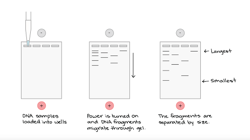
Gel Electrophoresis: Molecular Biology Science Activity | Exploratorium ... Prepare your gel: Make a 0.2% sodium bicarbonate buffer by dissolving 2 grams of baking soda in 1 liter of water. You will need approximately 100 milliliters per set up—half to make the gel and half to run your samples. Make a 1% gel solution by adding 0.5 g of agar-agar powder to 50 mL of sodium bicarbonate buffer. PDF Protocol 4: Gel Electrophoresis Teacher Version the Genome PROTOCOL 4: GEL ELECTROPHORESIS TEACHER VERSION ⎕ STEP 1 Set up and turn on the LONZA system and laptop so it is ready to go once the gels are loaded. ⎕ STEP 2 Open a fresh gel cassette package from the LONZA system and insert gel cassette into gel dock by sliding into place. Then remove white seals from gel cassette. Gel electrophoresis (article) | Khan Academy Gel electrophoresis is a technique used to separate DNA fragments according to their size. DNA samples are loaded into wells (indentations) at one end of a gel, and an electric current is applied to pull them through the gel. DNA fragments are negatively charged, so they move towards the positive electrode. Annotating A Gel | Get Your Science On Wiki | Fandom Part 1. Photo Editing: 1.Take your JPG or PNG file of your Gel and open it with a photo editing program (GIMP). 2. Under "Image" --> "Transform" rotate your picture by 90 degrees so that your wells are on top of the page. 3. Using the Crop tool Cut out the black borders leaving only the gel. 4.
Difference gel electrophoresis - Alchetron, the free social encyclopedia
How to Interpret DNA Gel Electrophoresis Results | GoldBio Agarose gel electrophoresis is a molecular biology method to analyze and separate DNA fragments based on their size. When you use gel electrophoresis to help you with molecular cloning, you may run into a common problem. For an example, you are ready to excise your digested plasmid DNA from agarose.
Gel electrophoresis (article) | Khan Academy
Gel Electrophoresis - Definition, Purpose and Steps | Biology Dictionary The broad steps involved in a common DNA gel electrophoresis protocol: 1. Preparing the samples for running The DNA is isolated and preprocessed (e.g. PCR, enzymatic digestion) and made up in solution with some basic blue dye to help visualize the movement of the sample through the gel. 2. An agarose TAE gel solution is prepared
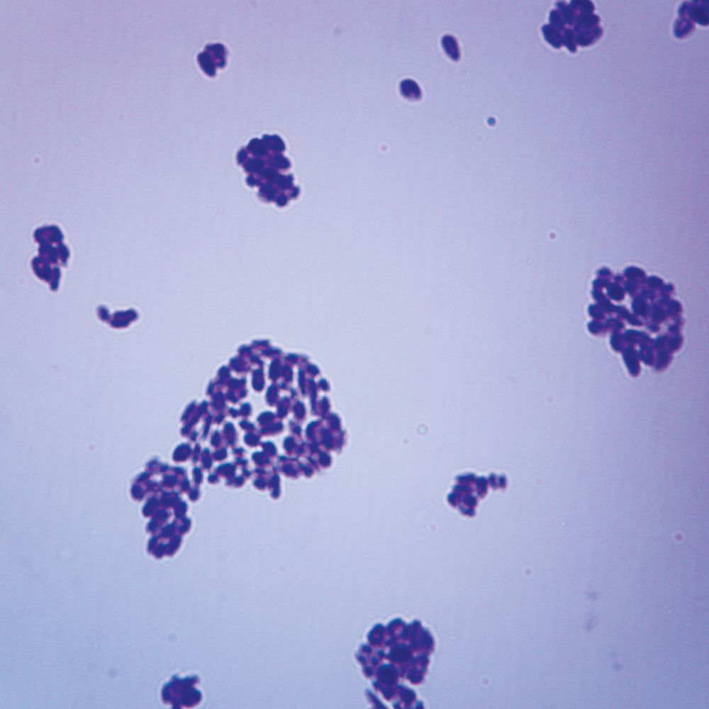
Saccharomyces Budding Cells, w.m. Microscope Slide | Carolina.com
Agarose Gel Electrophoresis: Results Analysis - Study.com Gel electrophoresis is a laboratory procedure used to separate biological molecules with an electrical current. Previously, we've discussed gel electrophoresis in the context of analyzing DNA....
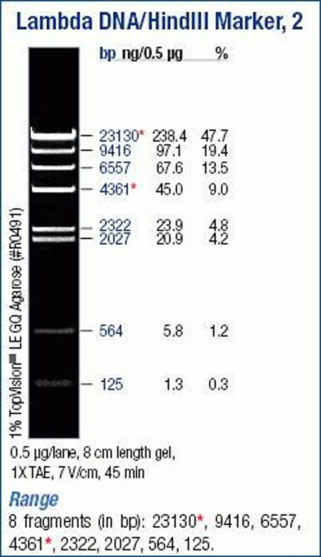
Lambda DNA/HindIII Marker - Thermo Fisher Scientific
Gel Electrophoresis - CSHL DNA Learning Center Gel Electrophoresis Description Transcript Keywords Info In the early days of DNA manipulation, DNA fragments were laboriously separated by gravity. In the 1970s, the powerful tool of DNA gel electrophoresis was developed. This process uses electricity to separate DNA fragments by size as they migrate through a gel matrix.
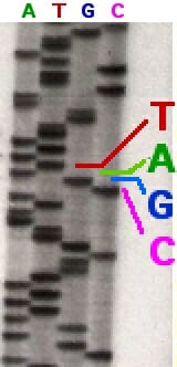
DNA Sequencing - Definition, Methods & Examples | Biology Dictionary
A Complete Guide for Analysing and Interpreting Gel Electrophoresis Results The image is captured under the UV transilluminator instead of the gel doc system to show you the effect of EtBr on the gel electrophoresis results. Here due to the re-use of a gel as well as the buffer, the EtBr is not properly spread into the gel. Further, the traces of the previous EtBr is also present in the gel.
Horizontal Electrophoresis | LSR | Bio-Rad
Agarose Gel Electrophoresis: Principle, Procedure, Results Agarose gel electrophoresis is a powerful separation method frequently used to analyze DNA fragments generated by restriction enzymes, and it is a convenient analytical method for separating DNA fragments of varying sizes ranging from 100 bp to 25 kb. DNA fragments smaller than 100 bp are more effectively separated using polyacrylamide gel ...

Animal Cell Model Diagram Project Parts Structure Labeled Coloring and ...
Agarose Gel Electrophoresis for the Separation of DNA Fragments Agarose gel electrophoresis is the most effective way of separating DNA fragments of varying sizes ranging from 100 bp to 25 kb 1. Agarose is isolated from the seaweed genera Gelidium and Gracilaria, and consists of repeated agarobiose (L- and D-galactose) subunits 2.
What would the RNA banding pattern look like if mRNA is subject to ...
E-Editor 2.0 Software | Thermo Fisher Scientific - US Just capture an image of the gel and use the E-Editor 2.02 software to: Align and arrange the lanes in the image Save the reconfigured image for further analysis Copy and paste selected lanes or the entire image into other applications for printing, saving, emailing, and/or publishing Analysis with E-Editor 2.0 is fast and convenient
Post a Comment for "42 labeled gel electrophoresis photo"