40 ear diagram
Ear Anatomy: Understanding the Outer, Middle, and Inner Parts of the Ear The external auditory meatus, or ear canal, is a narrow canal that leads from the concha to the tympanic membrane, or eardrum. Sound waves are delivered through this canal. This canal is prone to ear infections. Tragus This is the small, rigid part of the ears along the front of the ear, adjacent to the face. › human-body › earHuman Ear: Structure and Functions (With Diagram) ADVERTISEMENTS: In this article we will discuss about the structure and functions of human ear. Structure of Ear: Each ear consists of three portions: (i) External ear, ADVERTISEMENTS: (ii) Middle ear and (iii) Internal ear. 1. External Ear: It comprises a pinna, external auditory meatus (canal) & tympanic membrane. (i) Pinna: ADVERTISEMENTS: The pinna is […]
Ear Anatomy - Outer Ear | McGovern Medical School The outer ear is made up of cartilage and skin. There are three different parts to the outer ear; the tragus, helix and the lobule. EAR CANAL The ear canal starts at the outer ear and ends at the ear drum. The canal is approximately an inch in length. The skin of the ear canal is very sensitive to pain and pressure.

Ear diagram
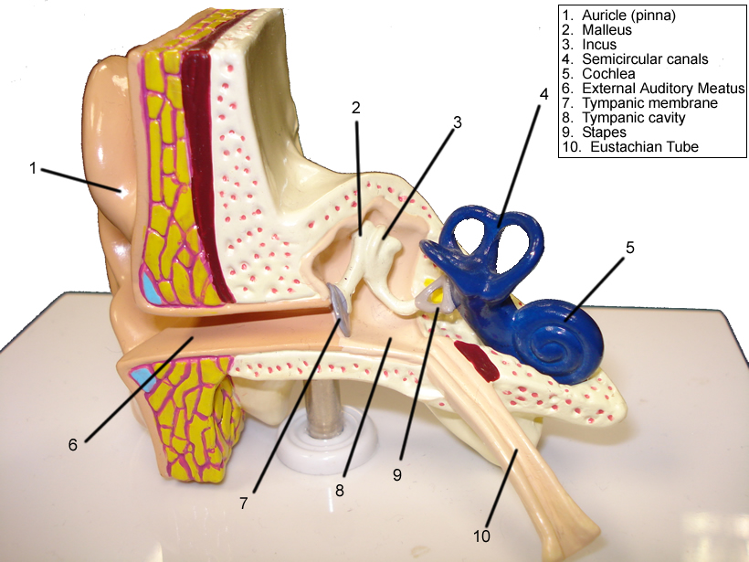
bodytomy.com › human-ear-diagramHuman Ear Diagram - Bodytomy One such organ is the ear that helps us in the process of hearing and balancing. The sound waves entering the ear get converted into electric impulses for the brain to understand and interpret. Let us take a look at the human ear structure with the help of a diagram, and understand its functions a little more closely. The Structure of Human Ear Anatomy of the Ear | Inner Ear | Middle Ear | Outer Ear The middle ear includes: eardrum. cavity (also called the tympanic cavity) ossicles (3 tiny bones that are attached) malleus (or hammer) - long handle attached to the eardrum. incus (or anvil) - the bridge bone between the malleus and the stapes. stapes (or stirrup) - the footplate; the smallest bone in the body. › human-body-maps › middle-earMiddle Ear Anatomy, Function & Diagram | Body Maps Jan 21, 2018 · Also known as the tympanic cavity, the middle ear is an air-filled, membrane-lined space located between the ear canal and the Eustachian tube, cochlea, and auditory nerve. The eardrum separates ...
Ear diagram. The Ear: Anatomy, Function, and Treatment - Verywell Health Essential organs of human hearing and balance, the ears are located on either side of the head, at the level of the nose. Separated into an inner, middle, and outer ear, each ear is an intricate and complicated mixture of bones, nerves, and muscles. Naturally, these structures are at the heart of hearing loss problems as well as those affecting ... Ear Diagram Vector Art, Icons, and Graphics for Free Download Browse 38 incredible Ear Diagram vectors, icons, clipart graphics, and backgrounds for royalty-free download from the creative contributors at Vecteezy! Ear anatomy: Parts and functions - Kenhub The ear is a complex part of an even more complex sensory system. It is situated bilaterally on the human skull, at the same level as the nose. The main functions of the ear are, of course, hearing, as well as constantly maintaining balance. The ear is anatomically divided into three portions: External ear Middle ear Internal ear Label Parts of the Human Ear - University of Dayton Label Parts of the Human Ear. Select One Auditory Canal Cochlea Cochlear Nerve Eustachian Tube Incus Malleus Oval Window Pinna Round Window Semicircular Canals Stapes Tympanic Membrane Vestibular Nerve. Select One Auditory Canal Cochlea Cochlear Nerve Eustachian Tube Incus Malleus Oval Window Pinna Round Window Semicircular Canals Stapes ...
Ear Anatomy, Diagram & Pictures | Body Maps - Healthline There are two main sections within the inner ear: the bony labyrinth and the membranous labyrinth. The cochlea, the hearing organ, is located inside the inner ear. The snail-like cochlea is made up... Ear Canal Diagram, Pictures & Anatomy | Body Maps Ear Canal Diagram, Pictures & Anatomy | Body Maps Human body Head Ear Ear canal External acoustic meatus The ear canal, also called the external acoustic meatus, is a passage comprised of bone and... Ear Anatomy, Diagram & Structure | What are the Parts of the Ear ... The following ear diagram depicts the inner ear, which contains sensory organs for hearing and balance, and the outer ear, which includes superficial structures. The middle ear is sandwiched... en.wikipedia.org › wiki › Ear_canalEar canal - Wikipedia The ear canal (external acoustic meatus, external auditory meatus, EAM) is a pathway running from the outer ear to the middle ear.The adult human ear canal extends from the pinna to the eardrum and is about 2.5 centimetres (1 in) in length and 0.7 centimetres (0.3 in) in diameter.
healthjade.com › human-earHuman Ear Anatomy - Parts of Ear Structure, Diagram and Ear ... Human ear The ear is divided into three anatomical regions: the external ear, the middle ear, and the internal ear (Figure 2). The external ear is the visible portion of the ear, and it collects and directs sound waves to the eardrum. The middle ear is a chamber located within the petrous portion of the temporal bone. Ear Diagram - Concha Audiology The cochlea is a fluid-filled organ essential for the transduction of mechanical (vibration) energy to electrical (nerve impulse) energy. Vibrations from the stapes on the oval window cause waves within the fluid, which causes the basilar membrane to move. The movement of the basilar membrane causes a shearing action of hair cells (outer and ... Ear Diagram | Worksheet | Education.com Ear Diagram. Open your ears for some interesting science! Your little biologist will learn the basic parts of the ear with this diagram. When he's finished reading, he'll enjoy a word search to review ear vocabulary. Inner Ear Diagram Photos and Premium High Res Pictures - Getty Images 119 Inner Ear Diagram Premium High Res Photos Browse 119 inner ear diagram stock photos and images available, or search for human ear to find more great stock photos and pictures. of 2 NEXT
Lyric hearing aid: a rare cause of benign necrotising otitis externa ...
Free Ear Diagram Templates - Edrawsoft Ear Diagram Template Download Template: Get EdrawMax Now! Free Download Popular Latest Flowchart Process Flowchart Workflow BPMN Cross-Functional Flowchart Data Flow Diagram EPC Fault Tree IDEF Diagram Org Chart Basic Org Chart Photo Org Chart Creative Org Chart Family Tree Genogram Network Rack Diagram Network Topology CCTV Network LDAP

Normal Sinus Anatomy MediVisuals Medical Illustration
PDF the diagram - Central Institute for the Deaf the diagram EAR HOW WE HEAR 1. Sound enters the ear. 2. The ear drum vibrates. 3. The bones in the middle ear move. 4. The fluid inside the cochlea moves. 5. The hair cells inside the cochlea vibrate. 6. The auditory nerve is activated. 7. The message is sent to the brain. Outer Ear Middle Ear Inner Ear

Auricular acupuncture map - Seasons of Balance Family Acupuncture LLC
Picture of the Ear: Ear Conditions and Treatments - WebMD The ear has external, middle, and inner portions. The outer ear is called the pinna and is made of ridged cartilage covered by skin. Sound funnels through the pinna into the external auditory...

Rig Diagram: Eric Clapton, Cream (1967) | Guitar.com | All Things Guitar
Blank ear diagrams and quizzes: The fastest way to learn - Kenhub Ear diagrams (labeled and unlabeled) Accelerate your learning with interactive quizzes Sources + Show all Ear anatomy overview Although it's not obvious to look at, the ear is anatomically divided into three portions: External (outer) ear Middle ear Inner ear As you can imagine, there's a lot of associated anatomy to learn for each portion!

Gentry, Teresa M / Anatomy Diagrams
Ear - Wikipedia In mammals, the ear is usually described as having three parts—the outer ear, the middle ear and the inner ear. The outer ear consists of the pinna and the ear canal. Since the outer ear is the only visible portion of the ear in most animals, the word "ear" often refers to the external part alone.
Eye and Ear Models
How to Draw Human Ear Diagram With Labelling #HumanEar Thanks for watching our Channel. how to draw internal structure of human ear,diagram of human ear for class 8,diagram of human ear with labelling,structure o...

Image | Radiopaedia.org
Ear Diagram (English} | CID Free Download CONTACT US. 825 S. Taylor Avenue Saint Louis, MO 63110. Toll free: 877.444.4574 Tel: 314.977.0132 Fax: 314.977.0023

Ear piercing Types – Tragus, antitragus, Industrial, Daith, Earlobe ...
healthmd.net › lymph-node-locationsLymph Node Locations - Neck, Groin, Ear, Diagram, Pictures ... Nov 01, 2012 · Lymph nodes are small organs, size of which measures 1-2 cm and is present throughout the body. Very often their size is small but it may develop and grow bigger than normal size when it is infected due to certain medical disorder. The main function of the lymph nodes is eliminating the dead cells of […]
Post a Comment for "40 ear diagram"